Laboratory of Neurobiology
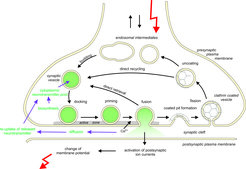
Neurons are the elementary units of our nervous system. They receive signals from other neurons, integrate them and then send them along axons to receiving neurons, often over long distances. Signal transduction between a sender and receptor neuron occurs at specialized structures, the synapses. Here, electrical signals are re-coded in chemical signals, the neurotransmitters. In the presynaptic compartment the neurotransmitters are synthesized and stored in small membrane vesicles, termed synaptic vesicles. Upon arrival of an electrical signal (termed action potential), voltage-gated calcium-channels located in the presynaptic plasma membrane open, leading to a transient increase in the intrasynaptic calcium concentration. This calcium-increase triggers the fusion of the vesicle with the plasma membrane, a process termed exocytosis. Neurotransmitters are released and diffuse towards the postsynaptic membrane, where they activate receptors and change the electrical properties of the receiving cell. On the presynaptic side, synaptic vesicles are retrieved by different endocytotic pathways and then, via intermediate steps, regenerated. Released neurotransmitters rapidly diffuse out of the synaptic cleft. They are rapidly sequestered by transporters residing in the plasma membrane of both neurons and adjacent glial cells.

Overview over the steps occurring during synaptic transmission between the arrival of an action potential in the presynaptic nerve terminal and the generation of an electrical signal in the postsynaptic cell.
Research of our group focuses on two major goals. First, we aim at arriving at a molecular description of vesicle docking and membrane fusion at the level of defined protein-protein and protein-membrane interactions and the associated conformational changes. In this regard, the SNARE proteins that function as fusion catalysts take center stage. We primarily use in-vitro approaches involving biochemical and biophysical methods, with the aim of isolating and characterizing partial reactions involved in the fusion pathway. In addition, we study fusion of endosomes which is less complex than the highly regulated neuronal exocytosis and thus more suitable for unravelling conserved molecular principles.
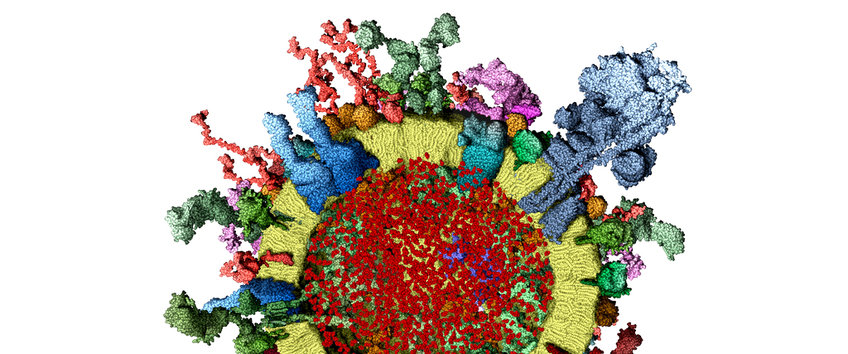
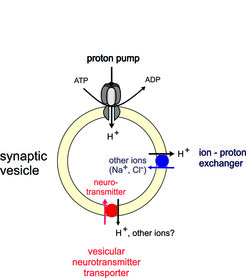
The second goal is aimed at arriving at a better understanding of the question how synaptic vesicles are filled within seconds with thousands of neurotransmitter molecules. This step is critical for neurotransmission but of all steps the least well understood. Several years ago we have developed a quantitative molecular model of synaptic vesicles, providing a solid foundation for quantitative work. Neurotransmitter uptake is mediated by specific transporters in the vesicle membrane that draw the energy for transport from an electrochemical proton gradient across the vesicle membrane. We want to understand how osmotic and charge balance is maintained during transport and how the transporters manage to remain operational while vesicular solute composition undergoes dramatic changes.
SNARE proteins – mechanism of action and regulation
During the past two decades, the key proteins mediating intracellular membrane fusions have been identified. The conserved core of the fusion machine includes the SNARE proteins that function as fusion catalysts, and SM- and CATCHR proteins that are involved in SNARE activation. Neuronal exocytosis involves at least two additional proteins, termed synaptotagmin and complexin (each represented by multiple isoforms) that are responsible for the tight regulation by Ca2+.
The molecular mechanism of regulated exocytosis is studied in many excellent laboratories. The field has been driven primarily by highly sophisticated physiological experiments in which individual proteins are being knocked out, replaced with mutant forms, or perturbed in other manners, resulting in a large body of data in which structural perturbations and neurotransmitter release are correlated. The biochemical foundation required to explain these experiments is still lagging behind. In particular, it has been difficult to reconcile the phenotypes with the emerging biochemical and physical properties of the proteins. Key questions such as the activation of SNAREs, the molecular roles of essential proteins such as Munc18 and Munc13, or the mechanism by which complexin controls late steps in the fusion reaction are still unclear.
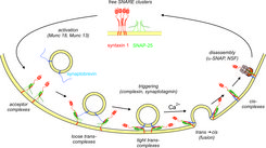
The neuronal SNAREs include syntaxin 1 and SNAP-25 at the synaptic plasma membrane and synaptobrevin (also referred to as VAMP) at the vesicle membrane. Like all SNAREs, these proteins undergo a regulated assembly disassembly cycle that constitutes the engine for membrane fusion and that is fueled by the AAA+-ATPase NSF in conjunction with its cofactor α-SNAP. Upon contact, the SNAREs associate in “trans” at the N-terminal ends of the SNARE motifs. A tight bundle of four α-helices is formed, each contributed by a different SNARE motif, which progresses towards the C-terminal membrane anchors (“zippering”), thus pulling the membranes tightly together and initiating fusion. Assembly is spontaneous and associated with a huge release of energy that is used to overcome the energy barrier for membrane fusion.

Intriguingly, members of the SNARE protein superfamily not only function in neuronal exocytosis but also in many additional intracellular fusion reactions. While in our work we mainly focus on the SNAREs responsible for neuronal exocytosis, we are also interested in the SNAREs mediating fusion of early and late endosomes. We have shown earlier that the structure of these SNARE complexes is almost identical to that of the neuronal SNARE complex. However, regulation of these SNAREs appears to be less complicated, making them good models for studying the basic features of SNARE function. We use these proteins not only for studying SNARE mechanisms but also to better understand intracellular targeting. Recently, in close collaboration with the group of Howard Shuman (University of Chicago), we discovered that certain pathogenic bacteria use SNARE-like proteins to hijack endosomal SNAREs. We found that these proteins can function as SNAREs but cannot be re-activated by NSF.
Our key goal is to precisely delineate the intermediate steps of the SNARE assembly and fusion pathway and to understand how the SNARE molecules are channeled along the pathway by regulatory proteins. Moreover, we want to understand at which state the proteins are arrested before the arrival of the calcium trigger, and how synaptotagmin, perhaps in cooperation with other proteins such as complexin, triggers exocytosis. To achieve these goals, we use “bottom-up” biochemical approaches. We express and purify SNAREs and accessory proteins, reconstitute them in artificial membranes, and then monitor vesicle docking and vesicle fusion using a variety of different approaches including both ensemble and single-vesicle assays.

Intermediate steps in SNARE-mediated membrane fusion, monitored by cryo-electron microscopy. Top: Crystal structure of the neuronal SNARE complex with transmembrane domains (from Stein et al. (2009) Nature 460, 525-528). Boxes indicate the position of mutations used in our experiments. Bottom: Cryo-EM images showing the intermediate stages during SNARE-mediated fusion of proteoliposomes (from Hernandez et al. (2012) Science 336, 1581-1584).
Vesicular uptake and storage of neurotransmitters

Loading and storage by synaptic vesicles of neurotransmitters is mediated by specific vesicular transporters that are coupled to an electrochemical proton gradient (generated by a V-ATPase) across the vesicle membrane. Since many years our group is interested in the mechanism by which the transporters for amino acid neurotransmitters (glutamate, GABA, glycine) and for ATP operate and how vesicular storage and refilling are regulated. Following our identification of the VGluTs many years ago, we use biochemical techniques for studying transport mechanisms. Examples of our approaches include functional reconstitution of purified transporters in artificial membranes, imaging of single vesicles, and measuring transport in purified synaptic vesicles whose interior is controlled by fusing them with liposomes containing defined solute compositions.


