Subnanometer Mechanics of Microtubule Dynamic Instability

Funding: DFG Research Training Group 2756 CYTAC
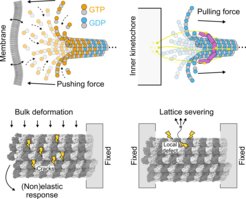
Eukaryotic microtubules (MTs) are cellular filaments that form the mitotic spindle, define the shape of axons and dendrites in neurons, and provide tracks for intracellular transport. MTs undergo stochastic switching between phases of growth and shrinkage driven by the hydrolysis of GTP nucleotides by tubulin dimers, building blocks that constitute the MT lattice. This semistable behavior of MTs, also known as dynamic instability, is crucial for MT function.

The detailed mechanism by which GTP binding and hydrolysis control the MT assembly and disassembly is still poorly understood, with two aspects standing out as essentially unresolved: (a) the missing link between GTP binding by tubulin and its conformation in solution, and (b) the mechanism of destabilization of the MT lattice by GTP hydrolysis.
Using a combined cryo-electron microscopy (cryo-EM) and molecular dynamics approach, the aim of the project is to characterize the mechanics and energetics of a single tubulin dimer depending on its nucleotide state as well as a complete MT complex in response to GTP hydrolysis.

