Single Molecule Ultrafast X-Ray Diffraction
Collaboration and financial support
Sebastian Westenhoff - Uppsala Universtiy, Department of Chemistry - BMC, Biochemistry
Philipe Maia - Uppsala University, Department of Cell and Molecular Biology, Molecular Biophysics
- Federal Ministry of Education and Research (BMBF) through the joint research project 05K20EGA Fluctuation XFEL, (2020-2024)
- Federal Ministry of Education and Research (BMBF) through the joint research project 05K2024 NT-RAC (2024-2028)
- Deutsche Forschungsgemeinschaft (DFG, German Research Foundation) - CRC 1456/1 - 432680300 Mathematics of Experiments, Project C02 Stochastic computed tomography: theory and algorithms for single-shot X-FEL imaging (2021-2024)
X-ray diffraction is the primary source for structure determination of biomolecules. Due to the low intensity of available x-ray sources, crystallized samples are required to yield sufficient scattering signal. However, crystallization is experimentally challenging and biases the molecular structure. Newly developed x-ray sources, such as the free electron laser XFEL in Hamburg, will provide high intense illumination sources, such that scattering experiments with single bio molecules become possible. In the experiment, a stream of hydrated single molecules enters the x-ray beam by e.g. applying electrospraying techniques. Due to the high photon flux of the incident beam, a single molecule undergoes severe radiation damage eventually leading to Coulomb explosion, however high beam intensity is required to register enough elastically scattered photons on the detector. Hence understanding the competition between elastic and inelastic scattering in those experiments is crucial for structure determination.
To investigate the applicability of this approach, we examine:
- How the immediate radiation damage influences the scattering signal
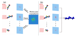
- How to reconstruct the molecular structure from multiple sparse and biased diffraction images
Radiation damage in single molecule X-ray scattering

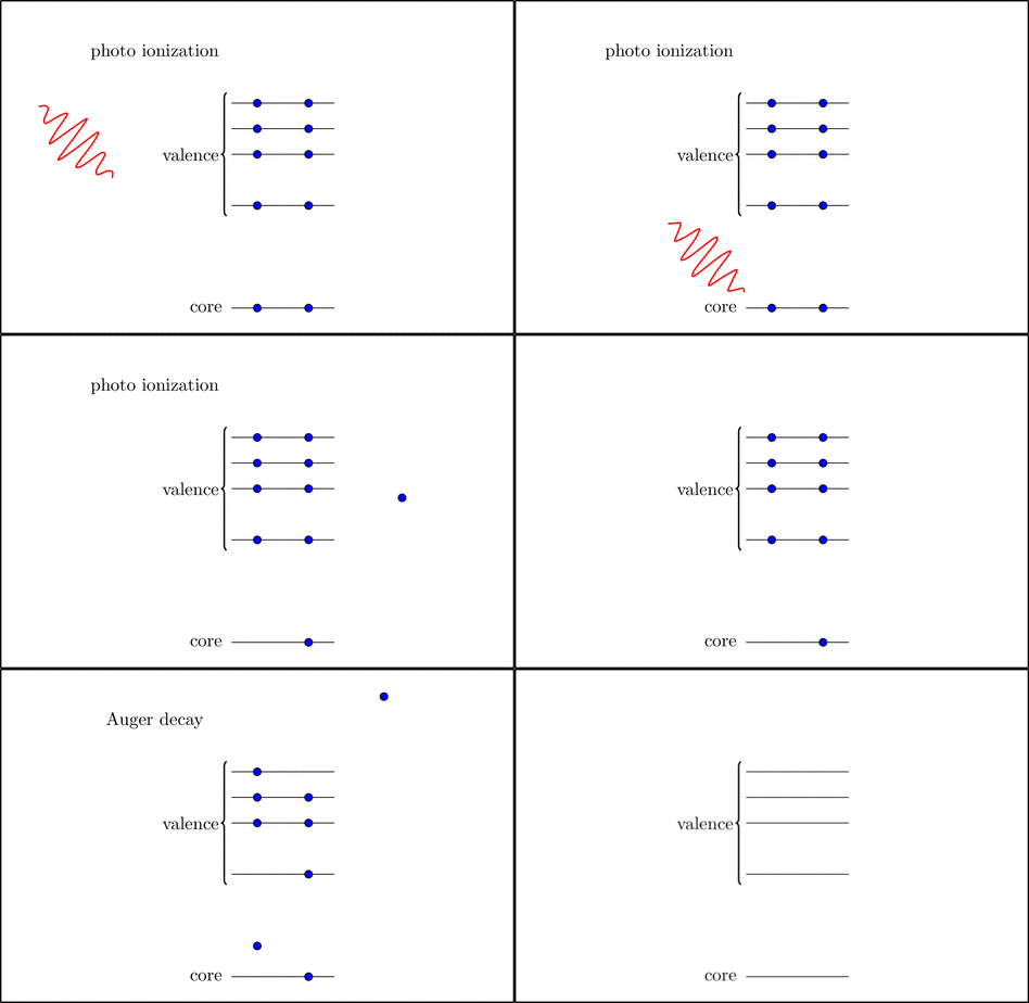
The primary process of radiation damage relevant here is photo ionization of electrons in the inner most atomic shell (K-shell). This is followed by Auger decay, during which an electron from an outer shell refills the core, while a second valence electron is emitted (see animation). Thereby the molecule charges up in sequences of K-shell ionization and Auger decay, which causes the deformation of the molecular structure (Coulomb explosion). To understand how fast structure desintegration proceeds within the recording time of the diffraction pattern, we study the two elementary processes, K-shell ionization and Auger decay in molecules via quantum chemical methods. Of particular relevance are electronic states with hollow K-shells, since ionization can outrun the refilling (Auger decay) of the K-shell, such that all electrons in the K-shell are stripped off [Inhester et al. 2012, Inhester et al. 2013].
Electron dynamics after core shell ionization
The Auger decay plays a crucial role in the radiation damage process during single molecule X-ray scattering experiments as the refilling of the K-shell hole opens the possibility for another photo ionization from the core shell, further accelerating the destruction of the target molecule by the Coulomb explosion. To better understand the impact of the refilling dynamics on the radiation damage we calculate the electron dynamics in space and time in atoms after sudden ionization from the K-shell or while coupled to ionizing X-ray radiation via the dipole approximation. To this end the time-dependent Schrödinger equation is solved using ab initio methods from quantum chemistry combined with problem adapted basis sets for the spatial representation of the wave function.
Structure determination from single molecule X-ray scattering

Reconstructing the structure from the experimental scattering images has to deal with two major problems of the setup. First, the orientation of the molecule during a single shot is unknown. Second, the number of photons scattered by one molecule is small and this leads to low signal to noise ratios. In this project, we therefore investigate new methods to retrieve the structure from the sparse scattering data with the help of statistical and analytical approaches.
- Bayesian orientation estimates and structure prediction
- Structure determination using three photons per image (photon correlations

