The Basic Paper: Real-Time MRI at a Resolution of 20 ms
We recently developed a unique method for real-time MRI that reduces image acquisition times to only 20 ms. It combines (i) FLASH MRI, which allows for rapid and continuous image acquisitions, with (ii) radial encoding, which offers motion robustness and moderate tolerance to data undersampling, and (iii) an iterative image reconstruction by regularized nonlinear inversion, which exploits the advantages of parallel imaging with multiple receive coils and enhances the undersampling by one order of magnitude.
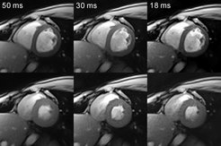
 Nonlinear inverse reconstructions with temporal and spatial filtering of diastolic and (bottom) systolic frames from real-time MRI movies of the human heart (2.0 mm in-plane resolution, 8 mm section thickness, TR = 2.0 ms) acquired from only 25 spokes (imaging time 50 ms), 15 spokes (30 ms), and 9 spokes (18 ms), respectively. The nonlinear inverse algorithm allows for a reconstruction with an undersampling factor of 20 as a fully sampled image with a 128 × 128 matrix would require 205 spokes.")
Real-time MRI refers to the acquisition, reconstruction, and display of magnetic resonance images without any unnecessary or even detectable delay. The method developed here realizes a temporal resolution of 20 to 30 ms for images with an in-plane resolution of 1.5 to 2.0 mm. It is based on a combination of fast low-angle shot (FLASH) MRI sequences with radial data sampling and image reconstruction by regularized nonlinear inversion. Corresponding iterative calculations are currently performed offline on a computer equipped with four graphical processing units after immediate real-time data export from the MRI system. Online reconstructions during scanning are accomplished by gridding using several combined datasets from successive acquisitions in combination with view sharing principles.
All measurements are carried out on an unmodified 3 T MRI system (TIM Trio, Siemens, Erlangen). For receive coils with a large number of elements (e.g, a 32-element cardiac coil) this process is supported by an online channel compression based on a principal component analysis. In general, the proposed real-time MRI method provides images free from motion artifacts, with a flexible and user-selectable trade-off between spatial and temporal resolution, and with access to different contrasts.
Although real-time MRI will cover a broad spectrum ranging from nonmedical studies of turbulent flow to the noninvasive monitoring of interventional (surgical) procedures, cardiovascular imaging, that is movies of the beating heart in real time, represents a first spectacular demonstration of the new capabilities and furthermore indicates a most important application which promises to benefit both patients and clinicians.
The new method visualizes cardiac functions at ultra-high temporal resolution (i.e., up to 50 frames per second) during free breathing and without the need for a synchronization to the electrocardiogram. Spoiled FLASH versions allow for the shortest possible repetition time (e.g., 2.0 ms) and – depending on the flip angle (power) of the radiofrequency excitation pulse – provide access to T1 contrast for dynamic contrast agent studies. Because of the very short echo times (e.g., 1.3 ms), the method does not suffer from off-resonance effects, so that the images neither exhibit susceptibility artifacts nor rely on fat suppression. On the other hand, versions with refocused or fully balanced gradients provide access to T1/T2 contrast. The choice of the gradient-echo time (in-phase vs opposed-phase conditions) may further alter the representation of water and fat signals and will allow for separate water/fat movies. Flow encoding (e.g., phase-contrast movies) is another option currently under investigation.
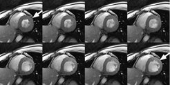
 with a measurement time of 33 ms per image and a spatial resolution of 1.5 mm (section thickness 8 mm). The eight successive images show the movement of the heart muscle of a healthy subject for a period of 264 ms during a single heartbeat. The images range from the systolic phase (top left: thickening and contraction of the left-ventricular myocardial wall) to the diastolic phase (bottom right: relaxation and expansion). The bright signal in the heart chambers is the blood.")
Apart from cardiac MRI other real-time applications deal with functional studies of joint movements (temporomandibular joint, knee) or address the coordinated dynamics of the lips, tongue, and soft palate during speaking (multi-lingual phonetics) or swallowing. Applications in interventional MRI, which refers to the monitoring of minimally invasive surgical procedures, are possible by interactively changing parameters such as image position and orientation.
For recent applications see Real-Time MRI in the applications section.