Research
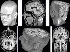
Methodologic advances in spatially resolved nuclear magnetic resonance (NMR) yield unique noninvasive insights into the anatomy, metabolism, and function of human organ systems. Extending structural magnetic resonance imaging (MRI), localized magnetic resonance spectroscopy (MRS) and functional MRI of the brain pave the way for new questions that range from basic neurobiologic research, e.g. involving transgenic animal models, to studies of human cognition.

As far as technical progress is concerned, already in 1985 we solved a key problem in MRI, i.e. long measuring times, by describing a new principle for the acquisition of rapid NMR images. The Fast Low-Angle SHot (FLASH) method revolutionized the scientific potential and clinical impact of MRI and strongly supported its commercial breakthrough as the leading modality in diagnostic imaging – with nowadays 100 million examinations per year worldwide.
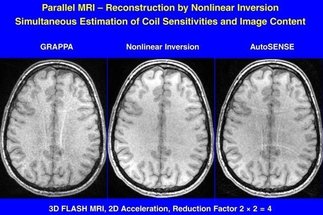
Our current research focusses on technical developments that further accelerate the MRI acquisition and reconstruction process toward real-time imaging and quantitative imaging. In particular, non-Cartesian (radial) encoding strategies and image reconstructions that solve a nonlinear inverse problem offer ways to exploit the speed gain achievable by extreme data undersampling. The ill-posed reconstruction problem benefits from temporal (or spatial) regularization to a preceding frame in a series of successive high-speed images.
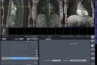
Real-time MRI studies not only overcome the motion sensitivity of classic MRI exams, but offer unprecedented access to arbitrary physiologic processes and body functions. For example, suitable applications in pediatric radiology now allow for studies of small children without the need for sedation or anesthesia.


