Unraveling the true identity of the brain of Carl Friedrich Gauss
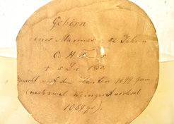
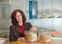
Walnut-like structures appear on the computer screen. They reveal what is inside the MRI scanner at the Biomedizinische NMR Forschungs GmbH: the 150-year-old brain of mathematician Carl Friedrich Gauss. Renate Schweizer monitors the MRI measurements as the images of the brain come into view section by section. Then she carefully places another brain on the examination table, where usually subjects are prepared for their MRI scans. It is the brain of Conrad Heinrich Fuchs – who was a medical scholar and founder of the University of Göttingen's collection of pathological specimens and, like Gauss, died in 1855. This unusual examination of historical brains from the collection at the Institute of Ethics and History of Medicine at the University Medical Center Göttingen has a specific purpose: "What scientists have recently been examining as Gauss' brain was not his brain at all, but actually originated from Fuchs. The two scientists' brains have been mixed up many years ago, and now need to be properly documented again," explains Schweizer, a biologist and psychologist, the surprising results of her investigations.
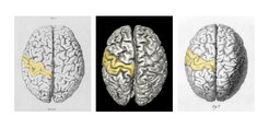
 and his copperplate of Gauss' brain from 1860 (right) show clear differences. The middle image is a recent MRI surface reconstruction of Gauss' brain. The central sulcus in the left half of each brain is highlighted in yellow.")
Schweizer made this unexpected discovery during extended anatomical studies in her own research field – the region of the brain around the so-called central sulcus. The gyrus on one side along the central sulcus processes somatosensory stimuli like touch, temperature, or pain, the gyrus on the other side controls muscular movements. Renate Schweizer suspected that Gauss' brain featured a very rare anatomical variation: a divided central sulcus. This variation is found in less than one percent of the population and remains without consequences to the people affected. If at all, it can cause minimal changes in motor and somatosensory function.
Images were not congruent
In the MRI surface reconstruction of the alleged Gauss brain, taken in 1998 by Jens Frahm’s team at the Biomedizinische NMR Forschungs GmbH, Schweizer had spotted this division of the central sulcus. To confirm her findings, she checked the original literature. Rudolf Wagner, a Göttingen anatomist and friend of Gauss, had not only preserved and studied the brains of both Gauss and Fuchs, but has also meticulously depicted the brain surfaces in his publications dating back to 1860 and 1862. But contrary to what she expected to see, Schweizer did not find the divided central sulcus in the images of Gauss' brain. Instead, her MRI surface reconstruction perfectly matched Wagner's picture of Fuchs' brain.

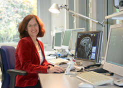
Renate Schweizer, neuroscientist at the Biomedizinische NMR Forschungs GmbH.
When Schweizer went to the collection at the Institute of Ethics and History of Medicine, her initial suspicion was convincingly confirmed: The the jar marked "C. F. Gauss" contained the brain taken from C. H. Fuchs, while the original brain taken from Gauss was in a jar marked "C. H. Fuchs". "My theory, according to the information I currently have, is that the brains were probably put into the wrong jars soon after Wagner's studies, at the time when the surface of the cerebral cortex was being re-evaluated by other scholars in 1864," the neuroscientist states. No further comparative studies of the brains of Gauss and Fuchs are known to have been undertaken afterwards and so no one noticed the mix-up. That the brains of Gauss and Fuchs are now properly assigned is also a significant finding for the Göttingen-based Gauss Society. "Its secretary, Axel Wittmann, provided excellent support right from the start of the project, and his extensive knowledge was extremely helpful in uncovering the – probably unintentional – mistake made so many years ago," Schweizer reports.
Invaluable treasure trove for science
The discovery also shows how important historical collections are for modern-day research. Schweizer confirms: "It is a stroke of luck that the brains in the collection are in very good condition and still accessible to researchers more than 150 years later." For the MRI measurements Schweizer closely collaborated with former team colleague Gunther Helms, an expert for investigations of brain specimens in the MR Research Unit at the Department of Cognitive Neurology at the University Medical Center Göttingen. As Jens Frahm, head of the Biomedizinische NMR Forschungs GmbH, emphasizes: "We are not looking for the genius in the gyri of the brain. Our aim is to fully document the historical specimens to preserve them for future research." All MRI scans, surface reconstructions, and photographs of the historic brains are now digitally archived, saving them as long-term scientific assets. But the MRI scans are already significant for present day research projects: Schweizer herself is currently using the images to gain novel insights into the divided central sulcus in Fuchs' brain below the surface of the cerebral cortex.
The new MRI results also demonstrate that earlier publications on the alleged Gauss brain did not provide false information. There, the mathematician's brain was described as normal. Walter Schulz-Schaeffer, head of the Prion and Dementia Research Unit of the Institute of Neuropathology at the University Medical Center Göttingen, inspected the recent images. He confirms that the brain of the brilliant mathematician and astronomer Gauss, like that of the physician Fuchs, is largely anatomically inconspicuous. The two brains are also similar in size and weight. "The age-related changes in Gauss' brain are normal for a 78-year old man. Changes in the basal ganglia could be indicative of high blood pressure," the neuropathologist comments.
Not every MRI scan of a historical specimen allows for such clear statements. That is why neuropathologists and MRI scientists are now cooperating to study how tissue and organs change if stored in alcohol for decades or centuries, and how MRI methods can be adapted to improve the interpretation of the images obtained – to make even better use of all the information stored in the historical specimens.
In the meantime the historical brains have again found their well-earned rest in the university collection – with no chance of a mix-up ever again. (cr/rs)

\".")