First light for our brand-new Olympus IXplore Spin system in the department
The Scope combines a dual camera spinning disc with a fast piezo stage, real time stitching and the suppression of unequal illumination artefacts. It’s mission: Automated 3D volume imaging of whole planarians at sub-cellular resolution. And boy, the neoblasts (planarian stem cells) really show up crisply :)
. A fluorescent in situ hybridization of the gene Piwi was performed to specifically label stem cells (red). The picture is an on-line stitched 2x5 mosaic composed of 10 individual images that were recorded with an Olympus IXplore Spin confocal spinning disc system at 20x magnification. Sample provided by James P. Cleland, MPI-BPC, Scale bar = 200µm")
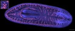
Confocal cross section of a fixed planarian Schmidtea mediterranea. Cell nuclei were stained with DAPI (blue). A fluorescent in situ hybridization of the gene Piwi was performed to specifically label stem cells (red). The picture is an on-line stitched 2x5 mosaic composed of 10 individual images that were recorded with an Olympus IXplore Spin confocal spinning disc system at 20x magnification. Sample provided by James P. Cleland, MPI-BPC, Scale bar = 200µm