Engineering nanobodies for super-resolution imaging and native protein complex isolation
Antibodies not only protect humans and other animals against disease-causing bacteria and viruses. They can also be used as tools for medical diagnostics and basic research. Conventional antibodies consist of light and heavy protein chains, and both are required for recognizing a target molecule (antigen). Alpacas, llamas, and camels, however, also possess simpler antibodies that lack light chains and bind to antigens via a single protein domain. Such domains can be produced in bacteria and are then called nanobodies. Compared to normal antibodies, nanobodies are ten-fold smaller, which is of great advantage in virtually all practical applications.
Researchers of the Department of Cellular Logistics headed by Dirk Görlich at the Max Planck Institute for Biophysical Chemistry made use of the recently established alpaca farm at the institute that by now houses ten animals. They immunized the animals with various antigens of the vertebrate nuclear pore complex (NPC) – a nanoscopic machine for transporting large biological molecules in and out of the cell’s nucleus – and eventually obtained a toolbox of nanobodies against the NPC. In the course of this work, however, they had to realize that currently available protocols for applying nanobodies in imaging and affinity purification suffered from substantial limitations.
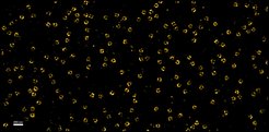
Nanobodies can be linked to fluorophores and then be used to stain sub-cellular structures so that they can be observed under a microscope. When fluorophores were attached in the traditional way, via the amino acid lysine, all tested nanobodies performed poorly in fluorescence microscopy – pointing to a systematic problem. Tino Pleiner and colleagues therefore explored an alternative, namely to label nanobodies via engineered surface cysteines. This was counterintuitive, because every nanobody already contains two internal cysteines and their modification would compromise the overall stability of the nanobody fold. However, the scientists found out that the labeling reaction is absolutely specific for the introduced surface cysteines when it is performed at low temperature. This strategy consistently yielded imaging reagents that could effectively position fluorophores as close as 1-2 nanometers to their antigens. Nanobodies labeled in this way are therefore ideal to exploit the full potential of super-resolution microscopy (Figure 1).

In contrast to conventional antibodies, which often bind disordered peptide motifs, nanobodies preferentially recognize structured regions of a protein or even contact multiple proteins of a protein complex at the same time. Therefore, it was much more difficult to identify where a nanobody actually binds. Interestingly, the engineered surface cysteines proved also useful as "position sensors" to report which region of an antigen a given nanobody binds. This was then revealed by crosslinking mass spectrometry.
Nanobodies are also used to purify protein complexes from crude cell extracts by affinity chromatography. Previously, nanobodies were chemically attached to an insoluble matrix, and bound protein complexes were released under conditions that destroy interactions between proteins. The Göttingen researchers now replaced the destructive step with a step that uses an enzyme to cut a bond and gently detach the nanobody (along with any bound protein complex) from the matrix. Bound protein complexes thus stay intact and can be studied further. In the future, this strategy can be applied to nanobodies that recognize tags commonly added to proteins (for example GFP) to isolate virtually any protein complex for functional assays or structural analyses. (tp/dg)


