Possible biomarker for Parkinson’s discovered using new MRI method
Certain metabolic products are suitable as indication for Parkinson’s disease. Researchers at the Max Planck Institute (MPI) for Multidisciplinary Sciences, the University Medical Center Göttingen (UMG), and the German Center for Neurodegenerative Diseases (DZNE) have used this insight in a first step to develop a new diagnostics approach. The prerequisite for the discovery is a method developed by Max Planck researcher Stefan Glöggler that specifically visualizes metabolic molecules in magnetic resonance imaging (MRI) by signal amplification.
The symptoms range from tremors and balance disorders to mental impairment and even dementia: Parkinson’s affects more than 6,1 million people worldwide. A newly developed method by Stefan Glöggler, a researcher at the MPI for Multidisciplinary Sciences and the Center for Biostructural Imaging at the UMG, raises hopes that Parkinson’s disease can be detected at an early stage using metabolic products.
The innovative technique allows to track individual metabolic molecules and their biochemical transformation in real time using MRI. If metabolic processes are altered, this may indicate disease. “In the future, our technique should help to detect such diseases earlier and to treat them in a more targeted manner,” the scientist explains. “To do this, we use a special form of hydrogen to modify metabolic molecules in such a way that their MRI signal increases more than 10,000-fold. This signal amplification is necessary to be able to observe the molecules specifically.”
How lactic acid indicates Parkinson’s
In their recently published study in the journal Chemistry Methods, the researchers headed by Glöggler focus on the body’s own metabolic molecule pyruvate. Pyruvate plays a central role in the energy metabolism of living cells and is degraded to lactic acid. In clinical MRI studies, scientists are already using it as a biomarker – meaning a biological indicator – for cancer. The research team now showed that pyruvate could also serve as a marker for Parkinson’s disease.

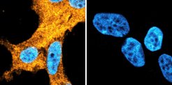
“In Parkinson's disease, the protein alpha-synuclein is abnormally altered. In the brains of those affected, it is formed in increased amounts, forming so-called fibrills that clump together and correlate to the impairment of nerve cells,” explains André Fischer, researcher at the DZNE and the UMG. His UMG colleague Felipe Opazo adds, “We have now demonstrated in a Parkinson’s cell model that cells that contain a lot of alpha-synuclein convert pyruvate into lactic acid twice as fast as cells that lack the protein. In the future, we hope to be able to detect Parkinson’s Disease early using the MRI technology established by the group of Dr. Glöggler.”
How protein structure affects the metabolism

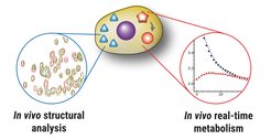
Thanks to Glöggler’s MRI method, the Göttingen scientists have also achieved another breakthrough, as Christian Griesinger, Director at the MPI for Multidisciplinary Natural Sciences, reports: “For the first time, we were able to simultaneously perform real-time metabolic analyses and determine protein structures in living cells in the same experiment. On the basis of a single cell sample, it is thus possible to find out whether metabolic disorders are present and to directly test whether errors in the structure of a specific protein – alpha-synuclein in the case of Parkinson's disease – are associated with them.” The interplay between changes in protein structures and altered metabolism is complex. “If we succeed in deciphering the influence one has on the other, this could lead to new insights into diseases and enable innovative therapeutic approaches,” says UMG researcher Tiago Outeiro.
Glöggler sees further advantages of his method in the fact that it is comparatively easy to use. “The technique is very fast and can even be combined with small, portable MRI devices,” the scientist summarizes. In subsequent experiments, the Göttingen researchers want to test whether their findings apply beyond the cell model to complex organisms. “This would bring us a big step closer to our goal of using our method in human medicine.” (sg/kf)


![[Fe]-hydrogenase catalysis visualized using para-hydrogen-enhanced nuclear magnetic resonance spectroscopy](/4868136/teaser-1734084319.jpg?t=eyJ3aWR0aCI6MzYwLCJoZWlnaHQiOjI0MCwiZml0IjoiY3JvcCIsImZpbGVfZXh0ZW5zaW9uIjoianBnIiwib2JqX2lkIjo0ODY4MTM2fQ%3D%3D--0262c0fa020d6ea4060dd5aacac2a1dcdee31120)


