Cholesterol/Lipids in the Pathology of Neurodegenerative Disorders
Primary defects in lipid metabolism are associated with myelin disease and vice versa, a growing number of neurodegenerative diseases, including multiple sclerosis (MS) and the hereditary leukodystrophy Pelizaeus–Merzbacher disease (PMD), are associated with a perturbed brain lipid metabolism. In the nervous system, the majority of lipids is found in myelin sheaths, a multilayered stack of membranes synthesized by oligodendrocytes. Myelinating oligodendrocytes contribute to axon integrity by providing trophic support and electrical insulation for impulse propagation. Myelination failure is hence associated with axon damage and dysfunction leading to deficits in cognition and motor abilities.
Multiple sclerosis (MS)
Multiple sclerosis (MS) is an inflammatory demyelinating disorder in which remyelination failure contributes to persistent disability.
We investigate the relationship between cholesterol and other lipids, myelination, and neurological parameters in mouse models of demyelination and remyelination. Using mouse models of MS and lipid-based therapies we dissect pathomechanisms and therapeutic strategies (Cholesterol recycling supports myelin repair - press release). However, endogenous repair strategies differ between the acute and chronic phases of myelin disease, and their efficacy attenuates with disease chronicity (Local cholesterol metabolism orchestrates remyelination: Trends in Neurosciences (cell.com)). Moreover, ketone bodies critically influence the CNS energy metabolism, especially during neuroinflammation (Von konservativ bis flexibel | Max-Planck-Institut für Multidisziplinäre Naturwissenschaften (mpg.de)).
In a wide range of neurological disorders including multiple sclerosis (MS) increased vascular permeability has been observed. In MS, the blood-brain barrier (BBB) permeability is increased in both newly forming demyelinating lesions and even in normal appearing white matter BBB disruption could potentially be secondary to pathology.

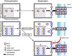
Fig. 1: Dietary cholesterol modulates the profile of growth factors, simultaneously enhancing OPC proliferation and oligodendrocyte differentiation, thereby facilitating remyelination and reducing axonal injury. Dietary cholesterol promotes repair of demyelinated lesions in the adult brain | Nature Communications

Fig. 2: Ketogenic diet (KD) supports repair after cuprizone mediated demyelination. Ketogenic diet ameliorates axonal defects and promotes myelination in Pelizaeus–Merzbacher disease | SpringerLink

Fig. 3: Cuprizone induces early BBB dysfunction and edema. A Experimental procedure to measure BBB permeability by quantifying extravasation of the tracers Evans blue (EB) and sodium fluorescein (NaFl), and edema (brain water). B The BBB is compromised already after a few days of cuprizone feeding, showing a predominant leakage in the corpus callosum (CC) in comparison to the cortex (Ctx). Blood-brain barrier hyperpermeability precedes demyelination in the cuprizone model | Acta Neuropathologica Communications | Full Text (biomedcentral.com)

Fig. 4: Resulting working model of the relationship between sterol metabolism in microglia/macrophages in an inflammatory demyelinating lesion and myelin repair. Microglia facilitate repair of demyelinated lesions via post-squalene sterol synthesis | Nature Neuroscience

Fig. 5: Neuronal cholesterol synthesis is essential for repair of chronically demyelinated lesions. While cholesterol recycling by CNS phagocytes orchestrates repair of acute lesions, repair after chronic demyelination is facilitated by neuronal cholesterol, especially by augmenting proliferation of oligodendrocyte precursor cells (Neuronal cholesterol synthesis is essential for repair of chronically demyelinated lesions in mice: Cell Reports).

Fig. 6 Cholesterol-dependent endogenous repair processes in demyelinated lesions. Working model of endogenous, cholesterol-dependent repair processes in acutely and chronically demyelinated lesions. (Local cholesterol metabolism orchestrates remyelination: Trends in Neurosciences (cell.com)).

Fig. 7 Proteome profiling of acutely isolated cortical cells reveals astrocytes are robustly equipped enzymes related to oxidative energy metabolism. Neurons metabolically benefit from feeding ketogenic diet during experimental neuroinflammation (Ketogenic diet uncovers differential metabolic plasticity of brain cells | Science Advances).
Pelizaeus–Merzbacher disease (PMD)
Pelizaeus–Merzbacher disease (PMD) is a leukodystrophy without therapeutic options. In most cases, PMD is caused by a duplication of the X-linked myelin gene PLP1 (proteolipid protein 1). In PMD oligodendrocytes, overexpressed PLP accumulates together with cholesterol which impairs the intracellular transport of proteins and lipids to the growing myelin sheath. In combination with the progressive loss of mutant oligodendrocytes, this leads to dysmyelination and demyelination in PMD. The MRI pattern of diffuse hypomyelination in PMD caused by PLP1 duplication is hence considered consequence of arrested brain maturation and lacks focal or inflammatory demyelination. Despite variable disease severity of PMD duplication patients, hypomyelination improves only to a minor extent over time. At later stages, axon degeneration is also a feature of the PMD pathology, leading to cortical atrophy that likely contributes to neurological impairment.
We hypothesize that the accessibility to the CNS determines the efficacy of lipid treatment. In the CNS, the different lipid classes target distinct aspects of the hypomyelinating pathology, i.e. the primary defect in myelin-forming oligodendrocytes and the subsequent damage of hypo/demyelinated axons. We aim to combine two important therapeutic targets, cholesterol for the support of remyelinating oligodendrocytes and ketone bodies for the metabolic support of the axonal compartment.
 rescues mitochondria enlargement and ameliorates impulse conduction in a mouse model of PMD (Plp1tgB).")
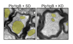
Fig. 1: Ketogenic diet (KD) rescues mitochondria enlargement and ameliorates impulse conduction in a mouse model of PMD (Plp1tgB).