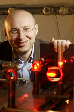
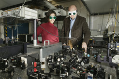
Stefan Hell and his research


© Max Planck Institute for Biophysical Chemistry


© Max Planck Institute for Biophysical Chemistry

© Max Planck Institute for Biophysical Chemistry

© Max Planck Institute for Biophysical Chemistry

© Max Planck Institute for Biophysical Chemistry

© Max Planck Institute for Biophysical Chemistry

© Max Planck Institute for Biophysical Chemistry

© Max Planck Institute for Biophysical Chemistry
. The image is based on recording 144 frames, the total image acquisition time was on the order of a second. Scale bar: 10 μm.")
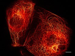
Live-cell imaging with parallelized RESOLFT nanoscopy. The image shows a RESOLFT recording of PtK2 cells expressing the fusion protein keratin 19–rsEGFP(N205S). The image is based on recording 144 frames, the total image acquisition time was on the order of a second. Scale bar: 10 μm.
© Andriy Chmyrov, Stefan Hell, Max Planck Institute for Biophysical Chemistry
, the STED image (right) shows considerably finer structures.")
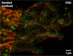
Two-color STED image of a glioblastoma, the most frequent malignant brain tumor in adults. Clathrin protein is green; ß-tubulin protein is stained red. In contrast to the blurred classical image (left), the STED image (right) shows considerably finer structures.
© J. Bückers, D. Wildanger, L. Kastrup, R. Medda; Max Planck Institute for Biophysical Chemistry
 provides approximately ten times sharper details of filament structures within a nerve cell compared to a conventional light microscope (outer image).")
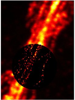
The STED microscopy (circular inset image) provides approximately ten times sharper details of filament structures within a nerve cell compared to a conventional light microscope (outer image).
© G. Donnert, S. W. Hell; Max Planck Institute for Biophysical Chemistry