Some of our research projects which involve animal experiments
Whenever possible, we replace animal experiments at our institute with alternatives such as cell cultures or computer simulations. However, these alternative methods are not always sufficient to understand complex biological processes in the organism. Therefore, animal experiments still remain indispensable in our research.
The welfare of our laboratory animals is of particular concern to us: We regard animal welfare, the best possible husbandry conditions, and responsible treatment of animals as our ethical obligation. In addition, animal welfare is an indispensable prerequisite for producing usable and reproducible scientific results.
You can find more information on the subject of animal experiments on our animal facility website as well as our portal page on animal welfare.
Below we present some projects from our research in which we use laboratory animals.
Research on the masters of regenerations – flatworms
Some flatworm species can literally be cut into tiny pieces and manage to regenerate complete and perfectly proportioned worms from each individual piece of tissue. In addition, many flatworm species do not seem to age and thus have an unlimited life span. These animals therefore provide a unique opportunity to study fundamental mechanisms of regeneration and tissue renewal. How do the remaining cells in a tissue “know” what is missing and needs to be replaced? What enables an animal to regrow lost body parts? Why do some species live seemingly forever, while others age and die?
The researchers hope to find answers to these questions using quantitative biology and comparisons between different species. To this end, they have assembled a large live collection of different flatworm species, representing a wide range of biological characteristics such as size, shape, regenerative abilities, and life expectancy.
Molecular switch influences regenerative ability
The aim of the research is to elucidate the molecular mechanisms of regeneration and tissue renewal and to understand how and why they change in evolution. Among other things, the scientists have already succeeded in identifying a crucial molecular switch, the so-called Wnt signal transduction pathway, which significantly influences the ability to regenerate. The flatworm Dendrocoelum lacteum cannot replace all missing body parts. However, when the researchers turned off the switch the animal was able to overcome its intrinsic regeneration defect.
Using mutant mice to understand neurological diseases

Diseases of the nervous system are diverse and can occur at any age. Some are rare, others, such as depression or Alzheimer’s disease, are widely spread. To understand how and why they develop, scientists often use mice. This is because genes and proteins from humans and mice that are relevant in neuroscience research differ only little, and the properties of mouse neurons closely resemble those of human neurons.
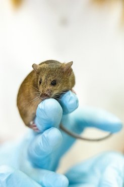
Research into neurological and psychiatric diseases often makes use of mutant mice into which, for example, foreign genes have been additionally introduced (‘transgenes’) or whose genes have been specifically modified (‘knock-in’) or rendered non-functional (‘knock-out’). Such methods make it possible to reproduce the genetic causes of corresponding diseases. Based on the changes caused by the inserted or knocked-out genes, scientists can draw conclusions about their mode of action and malfunction in diseases.
At the MPI-NAT, we also use mutant mice. Research on these animals allows us to gain a deeper understanding of the causes of neurological diseases and to contribute to the development of potential treatments. For example, our researchers succeeded in demonstrating that certain genetically caused malfunctions in signal transmission between nerve cells at so-called synapses can lead to brain diseases such as autism.
Alpacas as “development workers” in the production of antibodies
Antibodies protect us from disease. They are also an indispensable tool in science and medical diagnostics and could be used as medicines in the future.
“Common” antibodies recognize their antigens – for example, certain molecular structures of viruses or bacteria – with the help of two protein chains, the heavy chain and the light chain. However, alpacas and other camelids also produce simpler antibodies that can be reduced to so-called nanobodies by means of molecular biology techniques. When labeled with fluorescence or gold, such nanobodies can be applied to localize a protein of interest inside a cell with highest precision. They are also of interest for medicine: In 2021, our researchers produced mini-antibodies that have a neutralizing effect on the SARS-CoV-2 coronavirus even in the smallest quantities. The fact that they are very stable and can withstand extreme heat makes them a promising drug candidate to treat Covid-19.
Involvement of alpacas: vaccination and blood donation

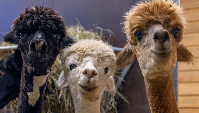
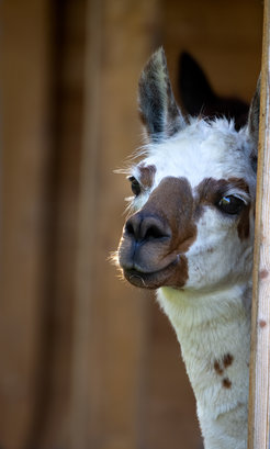
First, an alpaca is vaccinated with the antigen of interest, which is comparable to our autumnal influenza immunization. The animals hardly feel the tiny pinprick. The antigens are prepared in such a way that they are harmless to the alpaca. Its immune system then produces antibodies to fight the foreign body. About a quarter of a year later, a small amount of blood is taken from the alpaca from which scientists can isolate the building plans for these antibodies. Both, the inoculation and the quarterly blood donation only take a few minutes. After that, the animal has completed its task and can return to its herd in the pasture. In fact, the burden on an animal is so low that it can participate in several nanobody projects during its lifetime, while maintaining a good quality of life and a high life expectancy.
The actual work then takes place in the laboratory with the help of enzymes and bacteria. From the blood sample, which initially still contains blueprints for millions of different antibody variants, those that recognize the target molecule are sorted out. In the next step, bacteria produce the desired nanobodies in any quantity. Since the nanobody blueprints are known at this point in the process, production can be repeated at any time, and without the alpaca’s help. Another huge advantage: Many different nanobodies can be obtained from just one blood sample.
Husbandry with outdoor area and stable
Our 22 alpacas are kept on a spacious enclosure, which was designed by an expert in zoo design to be species-appropriate with pasture and gravel areas. They have access to their large stable at all times. The trusting animals can often be observed up close at the freely accessible enclosure next to our former Child Care Facility.
Nanobodies from Alpacas
New insights into sexual reproduction thanks to starfish and mice
A new life begins when an egg is fertilized by a sperm. In this fusion, the genetic information (DNA) from each parent is combined. The sperm and egg both contribute a single copy of each of their chromosome sets (in humans consisting of 23 chromosomes), which carry the DNA. The newly developing embryo thus inherits a complete set of 46 chromosomes. However, the oocyte – the egg’s precursor cell – contains two copies of every chromosome and must therefore lose half of these before fertilization can take place. This happens in a specialized cell division called meiosis.
In humans, this sensitive process is particularly error-prone compared to other mammals. If too many or too few chromosomes remain in the matured egg cell, there is a risk of miscarriage, infertility or diseases in the offspring such as Down syndrome.
Advantages of starfish research

To understand the complex physiology and development in humans, our scientists study this process in mice, but also in simpler species, including starfish. The oocytes of starfish are very large and transparent making them ideal for biochemical experiments and very easy to observe under the microscope. The eggs can be taken from the animals in large quantities as they mature outside the body in salt water. Since starfish go through a short development cycle and produce several thousand eggs, few animals are sufficient for research. The small wounds that occur in the adult individuals when the eggs are removed heal by the following day and are barely visible.
Modern mouse research provides valuable insights while using less animals

Mice resemble humans much more in their sexual reproduction than starfish. Their use in research helps to understand how errors occur during fertilization. To reduce the number of experimental animals, superovulation is induced in female mice. This involves injecting hormones that cause more eggs than normal to mature in the animals. The eggs then enter the fallopian tube during ovulation, where fertilization occurs. Similar hormone treatments are also used in women as part of fertility treatments. Research on mouse eggs already provided valuable insights into why fertility declines in women as they age.
In the meantime, oocytes from cattle and pigs are also used at the institute for many experiments in fertility research, since these are even more similar to humans in terms of oocyte development. Our researchers isolate these oocytes from ovaries obtained from slaughterhouse waste (without prior hormone stimulation).


