
In real-time
The FLASH 2 technology from the Biomedical NMR research group provides unprecedented insights into the body – and has great potential for clinical applications.
What does a heartbeat look like? How does our tongue move when we speak, swallow, or beatbox? Jens Frahm and his team can make it all visible. In 2010, with FLASH 2 (Fast Low Angle Shot), the researchers achieved a breakthrough in visualizing processes in the living body in real time. “For 25 years, we did not think it was possible. At least I did not,” Frahm admits today.

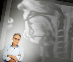
FLASH 2 uses a mathematical method for image reconstruction that requires only a few individual measurements per image. As a result, magnetic resonance imaging (MRI) is significantly accelerated. The measurement time for an image can be reduced by up to a hundredth of a second. “For 25 years, MRI has tried every trick in the book to eliminate the interference caused by motion. But motion is also information. For example, the heartbeat in cardiac arrhythmias, the swallowing process, or the movements of the gastrointestinal tract. 15 university hospitals are currently using FLASH 2 for research in these areas, including the Radcliffe Hospital at the University of Oxford (UK) and the Johns Hopkins University in Baltimore (USA). At the University Medical Center in Göttingen, Germany, four such devices are in use.
In recent years, the Biomedical NMR research group has also established national and international collaborations with scientists in the fields of musicology, speech therapy, and linguistics. Dirk Voit, a researcher in the group, explains: “There is no alternative to our technology for studying the speech apparatus and the pharynx. Until now, the only way to do this was to insert a camera through the nose or perform ultrasound on the throat. This did not work as well as real-time MRI.”
Child-friendly
 and Jens Frahm.")
FLASH 2 has another advantage for very young patients. “Real-time MRI is also ideal for pediatric radiology,” says Voit. “Radiologists have the problem that small children move around in the MRI and many measurements do not work. Conventional MRI scans often require children to be sedated or anesthetized. With a variant of real-time MRI, it is possible to image volumes – entire organs or the brain – with high image quality. “It takes one minute to completely measure an infant’s brain,” says Frahm. During this time, the child only needs to be held in position by one of the parents; anesthesia is no longer necessary. Less stress for the little patients, less work for the radiology staff. The Department of Pediatric Radiology at Leipzig University Hospital is already using real-time MRI with great success. “Our colleagues in Leipzig are phenomenal when it comes to clinical implementation and have done a lot of advertising for us, so that almost all German pediatric radiology departments are now contacting us,” says the research group leader. “Lübeck, Giessen, and Frankfurt are in the pipeline. The hospital in Philadelphia, perhaps the most important children’s hospital in the USA, is also interested.”
Hospitals also benefit from the comparatively low cost of FLASH 2, explains Frahm. “We have developed an algorithm on graphics cards that can calculate the images online. The computer with the graphics card can be connected to an existing MRI machine via a gigabit network cable. That is the whole installation – and you basically have a new MRI machine.”
How it all began


Paul Lauterbur developed MRI in 1973 and was awarded the Nobel Prize in Physiology or Medicine together with Peter Mansfield in 2003 for this technique. The procedure made it possible to take pictures of the inside of the body without using harmful X-rays. However, it was not yet suitable for clinical use. Although it could produce images of individual layers of the body, each slice took minutes to scan. An MRI scan therefore took several hours.
In 1985, with FLASH – the basis for FLASH 2 and real-time MRI – Frahm and his team succeeded in radically reducing the measurement time by a factor of 100. Today, FLASH is used worldwide for about 100 million examinations per year. The patent of Frahm and his colleagues is installed in virtually every hospital MRI scanner. It remains the Max Planck Society’s most successful patent to date and has already generated some 155 million euros in licensing income.
Star Trek medicine
In about 50 years, a technology that used to take hours has become a method for taking videos of the inside of the body – what does the future of MRI look like? “For example, we are currently developing volume technology. Then we will be able to combine the examination of individual organs into a kind of whole-body examination. All with one click,” says Frahm. Eventually, it will also be possible to follow minimally invasive procedures live inside the body using real-time MRI. “Theoretically, we already have all the prerequisites for a medical tricorder, just like in Star Trek.” If a body is in the appropriate magnetic field, it can be scanned with a handheld sensor, providing images and diagnostics. “This works with radio frequency antennas that pick up the MRI signals. We have already done that. So there is certainly a lot more to come in our field.” (kf)


