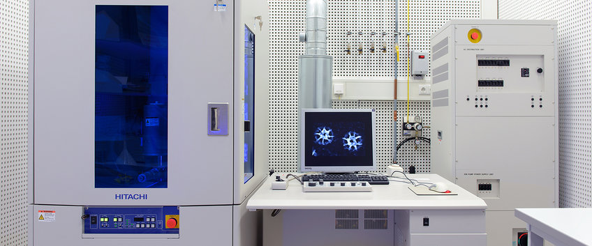
Facility for Field-Emission Scanning Electron Microscopy
The facility, with a background in the investigation of subcellular biological structures, provides support for the planning and conducting of SEM-based investigations. Following initial assessment of specimen suitability and the potential diagnostic conclusiveness of results potentially achievable by SEM, specimens for certain small-scale trial investigations can be prepared and evaluated by facility members upon request. More commonly, the facility offers a training for sample preparation for conventional SEM in general, or a more elaborate training for immuno-SEM of biological specimens. For large-scale, multi-sample studies, the facility additionally provides instructions for microscope usage and further training for specimen evaluation.
Major equipment
- Hitachi S5500. Scanning electron microscope, equipped with a cold field emission cathode and a special electron optical "in-lens" system. In addition to the secondary electron detector, the microscope is equipped with two backscattered electron detectors and one STEM detector. The S5500 is optimized for ultra-high resolution in the low nanometer range within small-area samples (< 20 mm2) at low accelerating voltages.
- Bal-Tec CPD 030. Critical point dryer, for gentle drying of biological specimens.
- Leica EM MED020. Highly versatile, high vacuum coating system that is equipped with a quartz-balance film thickness monitor and is currently used primarily for high resolution sputter-coating of specimens with thin layers of Pt or Cr metals.